A NIDA-funded brain imaging study has shown that regular users of marijuana have less gray matter than nonusers of the drug in the orbitofrontal cortex (OFC), a brain region that contributes to impulse control, decision-making, and learning. Such a deficit could make it more difficult to change counterproductive behaviors, including drug use.
The regular marijuana users’ OFCs also exhibited more functional and structural connectivity than those of nonusers. These differences could reflect an OFC response in the users to compensate for reduced volume by increasing coordination and efficiency of communication. However, the differences diminished with longer-term marijuana use, suggesting that the OFC’s capacity to offset relative gray matter deficits wanes after continued exposure to the drug.
Dr. Francesca Filbey of the University of Texas, who led the study, says that the differences she and her colleagues observed could have predated the study participants’ marijuana use. However, the researchers speculate that exposure to tetrahydrocannabinol (THC), the primary pharmacological agent in marijuana, may accelerate and exaggerate processes of neuronal pruning and neuron scaffolding that occur naturally as the brain matures.
Three Views of Cortex
For the study, Dr. Filbey and colleagues recruited 110 participants: 48 who reported using marijuana at least 4 times per week over the past 6 months, and 62 nonusers of the drug who matched the users in age and sex. All participants underwent magnetic resonance imaging (MRI) of gray matter and connectivity throughout the brain. Compared with nonusers, regular users of marijuana had:
- Less gray matter, an indicator of lower neuron density or volume, in two regions of the OFC (see Figure 1)
- More resting-state functional connectivity, indicating greater coordination of activity among neurons in the OFC network
- More organized myelinated axon bundles in the forceps minor, indicating more robust communication between the left and right OFC regions.
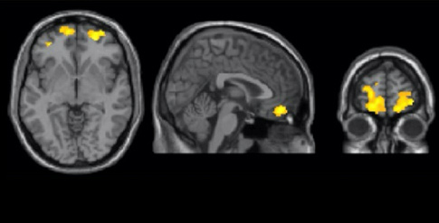
 Figure 1. Regular Marijuana Use Is Associated With Less Gray Matter in the Orbitofrontal Cortex (A) Yellow regions on MRI scans reveal areas in the orbitofrontal cortex (OFC) where regular users of marijuana had lower volume than did nonusers. (B) Regular users of marijuana, including those who used no other drugs, had lower gray matter density in the OFC than nonusers.
- Text Description of Figure 1
-
The figure shows a two-panel graphic. The top graphic, labeled “A” shows from left to right, a transverse, sagittal, and coronal MRI scan of the human brain, each of which having small regions (shown in yellow) in the OFC (in the frontal part of the brain) where regular users of marijuana had lower brain volumes than nonusers. The lower panel, labeled “B” shows a bar graph of gray matter density in the OFC (indicated on the vertical axis of the graph ranging from 0.30 to 0.50). As shown in the graph, nonusers had a gray matter density of about 0.45, regular users of marijuana of about 0.41, and exclusive, regular users of marijuana of about 0.40.
Less Gray, More White
The researchers hypothesize that the relative deficit in OFC gray matter in regular users of marijuana may help explain why they have a hard time stopping drug-related behaviors. Dr. Filbey says, “Our finding that chronic marijuana users had smaller gray matter volume in the OFC might manifest behaviorally, making it difficult for them to change learned behavior. For example, once someone has learned that using marijuana makes them feel good, it may be difficult for them to unlearn it and motivate themselves to change their behavior despite the negative consequences.”
Consistent with that idea, the researchers found that, among the regular users of marijuana in the study, less OFC gray matter correlated with higher scores on the 19-question Marijuana Problem Survey (MPS). The MPS assesses adverse psychological, social, occupational, and legal consequences of marijuana use, with higher scores indicating more problems. The finding suggests that regular users of marijuana with less OFC gray matter are more likely to persist in using the drug despite accumulating and worsening adverse experiences.
Dr. Filbey proposes that the more abundant communication and coordination among OFC neurons observed in regular users of marijuana may be an adaptation linked to their gray matter deficit. She says, “These increases in connectivity suggest a mechanism known as neuronal scaffolding, wherein more neurons are recruited to compensate for a decline in gray matter.” The study participants’ MPS scores support this interpretation. Among the regular users of marijuana, greater functional connectivity and thicker inter-OFC myelinated axon bundles each correlated with lower problem scores.
The effect of this compensatory response, if that is indeed what it is, appears to dissipate with prolonged use of the drug. Among the study participants, regular marijuana use was associated with rising OFC structural connectivity during the first 5 to 7 years of use and with falling structural connectivity thereafter (see Figure 2).
 Figure 2. Short- and Long-term Regular Marijuana Use Differentially Affects Structural Connectivity of the Orbitofrontal Cortex Fractional anisotropy, a measure of structural connectivity, increased between the two sides of the OFC for roughly the first 5 to 7 years of regular marijuana use, then declined with continued use.
- Text Description of Figure 2
-
The figure shows a scatter plot of fractional anisotropy in the OFC over more than 30 years of regular marijuana use. The vertical (y-) axis shows fractional anisotropy ranging from 0.50 to 0.65, and the horizontal axis shows years of regular marijuana use ranging from 0 to 36 years. The data points spread over almost the entire range of functional anisotropy, with a trend toward diminished anisotropy with longer marijuana use, indicated by a dashed line fitted to the data, showing a peak of fractional anisotropy at about 0.58 around 7 years of regular marijuana use and gradual decline to a fractional anisotropy of about 0.52 after more than 30 years of use.
Accelerated Pruning?
Dr. Filbey and her team didn’t delve into why regular users of marijuana differ from nonusers in their OFC gray matter and connections. However, they have a theory.
They suggest that marijuana exposure alters a natural process called synaptic pruning. During adolescence and young adulthood, the brain eliminates little-used neurons in the frontal cortex and wraps myelin sheaths around the axons connecting neurons that a person has relied on in dealing with the environment. Together, the two processes increase the region’s operating efficiency. Eliminating little-used neurons reduces the number of potential neuronal connections, but it also cuts interference between remaining neurons. Myelin sheaths increase the strength and rapidity of communication between neurons.
Dr. Filbey notes that the cannabinoid system regulates synaptic pruning. She says, “We speculate that with more cannabinoids in the system, this process may be accelerated.” Speeding up the process could produce the phenomena observed in the study—that is, users of marijuana with fewer OFC neurons and more OFC white matter than age-matched nonusers.
“However, future studies are needed to directly address this question,” Dr. Filbey says.
Searching for Definitive Answers
Although studies have consistently shown that marijuana changes brain function, studies have not agreed whether the drug alters brain structure. Inconsistent findings could reflect differences across studies in participant characteristics, such as amount and duration of marijuana use, age, sex, use of other substances, or psychiatric disorders. They also could arise from using different imaging methods or focusing on different brain areas. For example, studies that examine different brain regions might well produce contrasting findings. That’s because cannabinoid receptors, where marijuana binds and exerts its main pharmacological effects, are distributed unevenly throughout the brain.
Dr. Harold Gordon, a program official within NIDA’s Epidemiology Research Branch, says, “To compensate for discrepancies from prior studies due to subject characteristics and methodology differences, this study used a large, carefully controlled sample and imaged the entire brain with three imaging methods.”
Dr. Gordon says, “Since this was a cross-sectional study, it still remains to be determined whether the brain alterations preceded, or were a consequence of, cannabis use. Prospective studies, such as the Adolescent Brain Cognitive Development Study that will follow a large group of children over a 10-year period, would be needed to determine cause and effect. In addition, long-term studies following abstinence would be needed to determine whether the changes were reversible.”
Dr. Filbey agrees that more longitudinal research is needed to understand the effects of marijuana use. To help address questions about causality and to determine whether regular use of marijuana affects other processes, such as behavior, her group plans to continue to follow study participants over a number of years.
She says, “The next step for this research is to follow up with these individuals at different time points to see how their brains change over time in comparison to themselves, not a control group.”
This study was supported by NIH grant DA021632.
Source:
Filbey, F.M.; Aslan, S.; Calhoun, V.D.; et al. Long-term effects of marijuana use on the brain. Proceedings of the National Academy of Sciences 111(47):16913-16918, 2014. Full text