The brain, like an ocean, guards mysteries of the deep. Many of the structures and activities that make us what we are, mentally and emotionally, reside or occur far from the surface. Until now, researchers have observed deep-brain processes in living subjects only indirectly, through nuclear imaging or magnetic resonance techniques. Soon, however, if new technologies being developed by NIDA-funded scientists bear out their promise, investigators will have the means to view structures anywhere inside the organ and even activate or deactivate specific brain circuits at will. The technologies powerfully enhance neuroscientists' ability to understand how the brain functions—not only in drug abuse but across the spectrum of neurological disease. One of the new tools also opens the possibility of controlling neural circuits to counter neurological disease.
Wee But Mighty Scope
In an impressive feat of engineering, researchers have created a device about the size of a dime that performs the job of a powerful tabletop microscope. The new tool sends an optic fiber bundle measuring less than 1 millimeter in diameter into a test animal's brain. The fibers deliver light to the target site within the brain and then relay images to a packet of miniaturized external components so small that a mouse can carry it on its back.

This microendoscope, designed by Dr. Mark J. Schnitzer and colleagues at Stanford University's James H. Clark Center for Biomedical Engineering and Sciences, can provide clear images of structures up to about three-tenths of a millimeter away from the probe tip, which can reach a depth of 1 centimeter or so into the brain. The tool is more powerful, but has a thinner probe and smaller overall size, than earlier microendoscopes.
Dr. Schnitzer's team plans to use the technology to reveal critical neurological processes as they happen. For example, researchers might observe structural changes in the circuitry of the hippocampus during memory formation or monitor a tumor growing on the brain stem. "The technology will provide a great deal of information about how the living brain works at a very deep level—a scientific opportunity unavailable before," says Dr. Thomas Aigner of NIDA's Division of Basic Neuroscience and Behavioral Research.
Achieving Depth
A physicist who recently turned his attention to neuroscience, Dr. Schnitzer and his colleagues built the new technology on the foundation of fluorescence microscopy. In a well-established technique called one-photon fluorescence microscopy, researchers infuse tissue with a light-sensitive dye. Shining a bright light on the tissue causes the dye to emit photons that form an image. However, because brain tissue can scatter the photons before they reach the detector, the images lose resolution when the technique is applied more than about 100 micrometers deep. Consequently, researchers developed two-photon fluorescence imaging. Although this technique produced clear images just below the brain surface, it could only penetrate about 500 micrometers into the brain.
 scanning mirror. In this image, the structure is shown next to a dime. The microscope is sufficiently small that an adult mouse can carry the 2.9-gram device on its head.") Researchers Develop Miniature Microendoscope: The external components of a two-photon fluorescence microendoscope consist of a combination of tiny lenses, fiber-optic technology, and a microelectromechanical systems (MEMS) scanning mirror. In this image, the structure is shown next to a dime. The microscope is sufficiently small that an adult mouse can carry the 2.9-gram device on its head.
Researchers Develop Miniature Microendoscope: The external components of a two-photon fluorescence microendoscope consist of a combination of tiny lenses, fiber-optic technology, and a microelectromechanical systems (MEMS) scanning mirror. In this image, the structure is shown next to a dime. The microscope is sufficiently small that an adult mouse can carry the 2.9-gram device on its head.To achieve deeper visualization, Dr. Schnitzer and colleagues inserted an ultrathin probe—0.35 to 1.0 millimeter in diameter—through the skull and into the brain. The device's external components include microlenses and micromotors that power scanning, alignment, and focusing mechanisms.
The first portable two-photon fluorescence microendoscope weighed 4 grams and was the size of a matchbox. The team's latest version weighs only 2.9 grams and is about the size of a dime (see photo). In the external pack, a direct-current micromotor drives the focusing mechanism and controls silicon mirrors smaller than one square millimeter.
In one test with anesthetized mice, the 4-gram microendoscope produced detailed images of blood vessels located in the hippocampus, a structure located near the core of the animal's peanut-sized brain. In an initial test of the optical capabilities of the smaller microendoscope, the team imaged single grains of pollen and more recently microvasculature. That resolution is sufficient for portraying cells within the brain.
"Other teams are starting to use our probe technology for deep brain imaging in anesthetized animals," says Dr. Schnitzer. "My colleagues and I are developing microendoscopes that can image the brains of awake animals. We believe that scientists will find many uses for these devices in their research."
Even more valuable would be imaging technology that visualizes an animal's brain over weeks, months, or longer. Such a tool would have wide applicability in neuroscience, especially in the study of neural circuit development and cellular changes in response to experience. "Our goal is to build a general imaging tool that scientists can use to study the brain for a protracted period of time," Dr. Schnitzer says.
Ultimately, scientists may also combine two-photon fluorescence microendoscopy with genetic techniques to view particular neural circuits in active animals.
Remote Control of Brain Cells
Dr. Karl Deisseroth of Stanford University led the team that has developed optical remote control of discrete neural circuits. With the new technology, researchers activate highly selective sets of neurons in an animal's brain by shining a blue light on them. To deactivate the neurons, the researchers hit them with an amber light.
In the most dramatic demonstration of the optical remote to date, the researchers twitched rodents' whiskers by directing blue light at the controlling neurons within the animals' motor cortex (see box below). In previous trials, they made worms (Caenorhabditis elegans) wriggle and become still by alternately exposing them to blue and amber light. (To see a video of this experiment, visit http://www.nature.com/nature/videoarchive/braincellonoffswitch/).
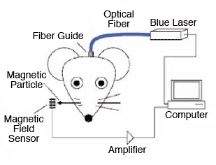
Blue Light in Brain Twitches Rodent Whiskers

Stanford University researchers used their optical remote to activate neurons in the brains of rats and mice. Dr. Karl Deisseroth and colleagues targeted neurons in a layer of the motor cortex that controls movement of a whisker. First, the scientists introduced a gene into the neurons that made them produce the light-sensitive protein channelrhodopsin-2 (ChR2). Next, the investigators directed an optical fiber 200 μm in diameter to the cells and illuminated the protein with a blue laser diode. In response, positive ions rushed into the neurons, the cells fired, and the whisker moved. The diagrams show the optical-neural interface for cell stimulation and the magnetic field sensor used to measure the movements of the whisker, to which a magnetic particle had been attached.
Source
Journal of Neural Engineering 4:S143-S156, 2007.
Two proteins are keys to Dr. Deisseroth's system, which he developed in collaboration with his graduate student Feng Zhang, other colleagues in their laboratory at Stanford University, and collaborators at the Max Planck Institute of Biophysics, the Johann Wolfgang Goethe Institute, and the University of Wuerzburg in Germany. To make cells responsive to the "on" and "off" light signals, the researchers introduce DNA for the blue light-reactive protein channelrhodopsin 2 (ChR2) and the amber light-reactive protein Natronomonas pharaonis halorhodopsin (NpHR) into the cells they will target. For example, the rodents whose whiskers twitched carried DNA for these proteins in their cortical motor neurons, and the worm that moved and halted in response to colored light carried it in its muscle walls and motor neurons. Cells with this DNA manufacture the proteins and store them in their membrane surfaces.
The Stanford team's system brings a long quest to a successful conclusion. ChR2 and NpHR belong to the class of proteins called opsins, which biophysicists had for years sought to utilize as neural switches. When an opsin in a cellular membrane is exposed to light, it changes shape, allowing ions to flow into the organism. Shining a blue light on a cell that features ChR2 opens a molecular gate, and positive sodium ions rush in, generating action potentials. Shining an amber light on NpHR precipitates an influx of negative chloride ions that silences the cell. The natural sources of opsins are some single-celled organisms, which rely on the proteins to regulate their activities or internal processes. The researchers culled the DNA for ChR2 from a green algae, Chlamydomonas reinhardtii, and the source of DNA for NpHR is a bacteria-like organism called N. pharaonis.
"As a research tool, optical remote control of neurons represents a huge leap forward for scientists who explore neural circuitry and neural circuit dynamics," says Dr. Geraline Lin of NIDA's Division of Basic Neuroscience and Behavioral Research.
Finer Control
The optical remote represents a marked advance over brain stimulation by electrodes, which has been the mainstay of neurological research for more than a century. The optical filament that the researchers insert into the brain to deliver laser light to cells is much finer than a standard electrophysiology electrode (roughly the difference between the diameter of a hair versus that of a sewing needle) and so disrupts less brain tissue. The optical system can turn cells on and off at millisecond intervals, compared with second-long intervals with electrodes. Scientists can choose which cells to make light-reactive with opsin DNA and use the optical remote to zero in on specific types of neurons. Electrodes, in contrast, are much less selective; they stimulate all the cells in a region.
With these advantages, the optical remote has wide potential application for addiction research as well as basic neuroscience. "This tool can help scientists see how neural circuits go awry in drug abuse and other brain disorders, information that may ultimately translate into therapeutic strategies," says Dr. Deisseroth, a psychiatrist with expertise in bioengineering.
NIDA's Dr. Lin says that combining the optical technology with other existing investigative techniques offers unprecedented opportunities to advance understanding of addiction. For example, researchers might document the neural activity in a particular circuit of an animal's brain during drug withdrawal, then test whether producing the same activity through use of the optical remote would also cause withdrawal symptoms. If it did, the scientists would know that the circuit was instrumental in withdrawal, and the next step might be to test whether suppressing the activity—again using the optical remote—could alleviate withdrawal.
"Before using the optical remote in people, we must proceed carefully with all the appropriate testing," notes Dr. Deisseroth. "Such an implant is somewhat invasive, but no more than current deep-brainstimulation devices, such as the electrical pacemakers that therapeutically stimulate areas deep in the brain of Parkinson's patients.
Because optical remote technology requires that neurons produce opsins, the technique would necessitate the introduction of genetic material into human cells. Daunting as that may sound, medical scientists are already employing such techniques. For example, the U.S. Food and Drug Administration has approved studies in which researchers use a virus to introduce genes into the human brain as a potential therapy for Parkinson's disease.
The team led by Dr. Deisseroth is disseminating the optical remote technology and has provided the genetic tools and technical support upon request to scientists exploring a variety of neuroscience questions, including how fish move, how neuromodulators control neural circuit function in the mammalian brain, and how seizures begin and terminate.
For their part, Dr. Deisseroth and colleagues are currently using their invention to elucidate the causes of narcolepsy. In one experiment, they stimulated cells in sleeping mice that release the neuropeptide orexin (also called hypocretin) in the lateral area of the hypothalamus of sleeping mice. The animals awoke, verifying that these neurons, which are deficient in people with narcolepsy, regulate wakefulness.
"I believe this breakthrough technology will spur an enormous amount of research that examines and elucidates neural circuit activity dynamics under normal and disease conditions. It will also enable researchers to develop strategies to normalize the perturbed neural circuit activities," Dr. Lin says. "The research will ultimately translate into help for patients with various psychiatric diseases, including drug addiction, and neurological disorders."
Sources
Adamantidis, A.R., et al. Neural substrates of awakening probed with optogenetic control of hypocretin neurons. Nature 450(7168):420-424, 2007. [Abstract]
Zhang, F., et al. Multimodal fast optical interrogation of neural circuitry. Nature 446(7136):633-639, 2007. [Full Text (PDF, 1.7MB)]
Deisseroth, K., et al. Next-generation optical technologies for illuminating genetically targeted brain circuits. The Journal of Neuroscience 26(41):10380- 10386, 2006. [Full Text (PDF, 235KB)]
Piyawattanametha, W., et al. Fast-scanning two-photon fluorescence imaging based on a microelectromechanical systems two-dimensional scanning mirror. Optics Letters 31(13):2018-2020, 2006. [Abstract]
Flusberg, B.A., et al. In vivo brain imaging using a portable 3.9 gram two-photon fluorescence microendoscope. Optics Letters 30(17):2272-2274, 2005. [Abstract]